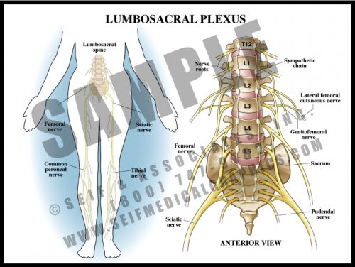
- Analogous to the brachial plexus, the lumbosacral plexus is a series of nerve convergences and separations which ultimately combine into several large terminal nerves.
- Plexi form a protective mechanism in that if one nerve root is damaged, a particular muscle might be weakened, but function would not be completely lost.
- The terminal nerves in the legs generally follow the course of the deep vasculature.
- Terminal sensory nerves to the feet are particularly vulnerable to diabetes, resulting in peripheral diabetic neuropathy. This frequently contributes to foot infections and the need for amputation.
-
-
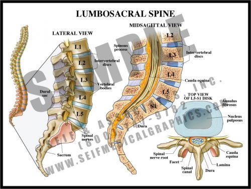
- The lumbar spine is composed of large, strong bones which must support the entire weight of the spine and head.
- The vertebral bodies are separated by fibrous discs which serve as shock absorbers. The discs have a fibrous ring (annulus fibrosis) and a gel-like center (nucleus pulposus).
- The spinal canal is formed by the pedicles, laminae, and the vertebral bodies and discs; the canal protects the distal portion of the axial nervous system, the cauda equina.
- The large transverse and spinous processes serve as support for the many paraspinous muscles which allow for the fine movement of the spine.
- The sacrum is the large, wedge-shaped bone forming the posterior part of the pelvic bowl, and is composed of fused vertebrae.
-
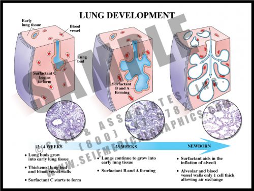
- The main reason that preterm infants are considered high risk is because their lungs are immature.
- Lungs develop as the airways bud and branch into an anlage of mesenchymal cells. Since respiration requires oxygen and carbon dioxide to cross over two layers of tissue (alveolar wall and capillary wall), these relatively thick-walled airways in preterm babies permit little gas exchange. High-pressure ventilation is required to assist the infant, and this pressure frequently results in the development of chronic lung disease (bronchopulmonary dysplasia).
- In addition, there are too few alveoli present for efficient oxygen supply until 2-3 weeks prior to term. Lungs continue to grow and develop new alveoli for several years after birth.
-
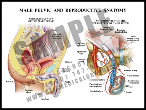
- The male pelvis contains the bladder and rectum, along with the internal portions of the reproductive system: the prostate, seminal vesicles, and the intrapelvic portions of the ductal apparatus.
- Sperm is produced in the testes, which lie in the scrotal sac outside of the pelvis. The sperm travels up the spermatic duct and is stored in the seminal vesicles. At ejaculation, sperm is released along with prostatic fluid, both of which travel down the urethra.
- The urethra has three parts: the prostatic portion; the membranous portion which passes through the urogenital diaphragm, and the penile portion.
- The spermatic cord contains a venous plexus, the spermatic artery and the spermatic duct.
-
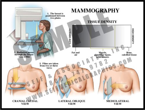
- Mammography is an imaging technique which allows visualization of breast tissue. Fat, glandular tissue and ductal tissue have characteristic densities and patterns.
- Solid tumors are generally easier to see in older patients with larger amounts of fat within the breasts, but can be difficult to see in patients with “dense” breasts (young women and women with fibrocystic breasts).
- Approximately 75% of breast cancers can be seen on mammography, with the patient’s breast characteristics being the largest determining factor.
-
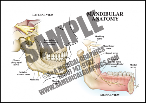
- The mandible, or lower jaw, is the bone that hinges to the skull and, together with the maxilla, forms the mouth.
- The mandibular nerve, the third and largest branch of the trigeminal nerve, runs along the mandible.
- The mandibular nerve has both sensory and motor functions. It divides into trunks and smaller branches to innervate the teeth and gums of the mandible, the lower lip and lower part of the face, and the muscles of mastication.
-
- This 2D dissolve animation shows how this maneuver can release shoulder dystocia impaction and allow for delivery of the fetal shoulder.
- Each animation has pause and play buttons to allow for more interactivity during viewing.
-
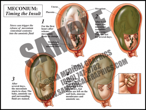
- Meconium is the dark, tarry contents of the fetal gastrointestinal tract. Meconium passage at labor and delivery is common and not a sign of fetal distress unless it is accompanied by other ominous signs.
- Meconium release into the amniotic fluid is however considered a sign of intrauterine fetal distress. This can be caused by placental insufficiency, maternal hypertension, and preeclampsia, among other things.
- When meconium is freshly released, it is clumpy and oats in the amniotic fluid. Within hours, it distributes particulate matter throughout the sac. Macrophages in the fetal skin, mucus membranes, and the amniotic sac phagocytize the particles, giving a green cast to the tissues.
- After several days, the fluid is greenish-brown but clear, and the fetus and membranes are stained. After several weeks, the fetal skin and fluid will clear, but the amniotic membranes, fetal mucus membranes, and nail beds remain stained.
-
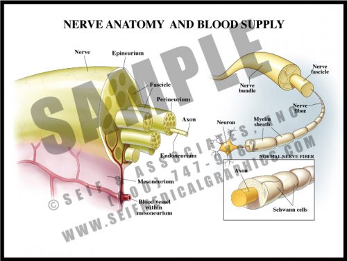
- Nerves are composed of multiple fibers which in turn are composed of many, many axons. Each successive grouping of fibers is surrounded by connective tissue.
- Axons are the long branches of nerves cells which transmit electrochemical impulses from one nerve to another or to and from target organs.
- The axons are surrounded by myelin, a kind of insulating material formed by oligodendrocytes. In diseases in which myelin is destroyed, electrochemical conduction is impossible, and even if the nerve fibers do not die, they are ineffective.
- Blood vessels travel through the mesoneurium and then divide into capillaries within the nerve to deliver oxygen and nutrients.
- Nerves are vulnerable to trauma, reduced oxygen levels, and diabetes.
-
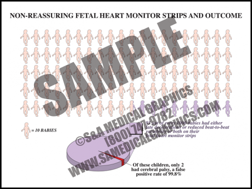
- One risk of fetal heart monitoring technology is false-positive results.
- In fact, “nonreassuring” FHM strips, or those which show either late decelerations, or reduced beat-to-beat variability, or both, have a greater than 99% false-positive rate in predicting cerebral palsy.
-
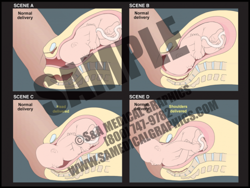
- This 2D animation demonstrates the nor- mal course of labor, showing the dilation of the cervix, descent of the fetus, delivery of the fetal head, and finally delivery of the fetal shoulders and body.
- This animation has pause and play buttons to allow for more interactivity during viewing.
-
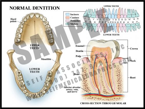
- There are normally 32 teeth, divided into 4 categories: molars, premolars, canines, and incisors.
- The teeth are embedded in the bones of the maxilla (upper jaw) and mandible (lower jaw) and are held in position by periodontal ligaments.
- The third molars (teeth #1, 16, 17, 32) are often vestigial and/or impacted. These are commonly known as “wisdom teeth”.
- The roots of the teeth are anchored within the bone and contain an artery, vein, and nerve which travel to the main portion of the tooth and divide within the pulp.
- Dentin covers the pulp and very hard enamel covers the dentin; the bone is covered with a mucosal tissue, the gingiva.
-
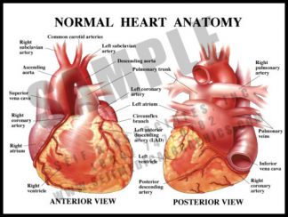
- Heart muscle is supplied by the coronary arteries, not by the blood flowing through the heart.
- The major coronary vessels are the right coronary artery (RCA) and left main coronary artery (LCA), both of which come directly off of the aorta via the coronary ostia.
- The LCA divides into the left anterior descending artery(LAD) and circumflex artery.
- The RCA has no major branches, and terminates as the posterior descending artery (PDA).
- There are may be variations in the anatomy.
-
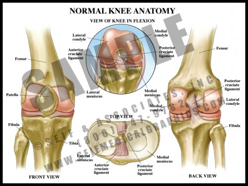
- One of the major joints in the body, the knee is required to support huge and repeated pressures over the course of a lifetime. The articular surfaces of the joint are covered with glassy-smooth articular cartilage, and the menisci act as shock absorbers with each step.
- The patella is a large sesamoid bone which lies within the quadriceps tendon. It articulates with the condyles of the femur.
- The anterior and posterior cruciate ligaments are located at the center of the joint and allow some rotatory motion. The lateral and medial collateral ligaments are attached on either side of the joint to maintain stability.
-
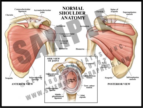
- The tendons of the deep shoulder muscles (infraspinatus, supraspinatus, and teres minor) conjoin with the shoulder joint capsule to form the rotator cuff.
- Rotator cuff injuries are common and may be difficult to treat.
- The head of the humerus rests in the glenoid fossa, a relatively shallow depression in the scapula. The rotator cuff holds the head in the fossa during movement.
- The shoulder joint is prone to dislocation due to the shallowness of the glenoid fossa.
- The acromioclavicular joint lies over the shoulder and sometimes develops fibrosis, making movement painful or stiff.
-
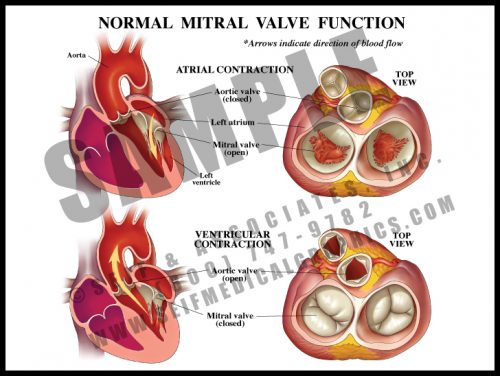
- Also known as the left atrioventricular valve, the mitral valve has 2 leaflets which are anchored to the ventricle floor by papillary muscles and chordae tendinae, as are the leaflets of the right atrioventricular valve (tricuspid valve).
- The aortic and pulmonary (pulmonic) valves are semilunar valves, and have thin cusps with thickened edges which seal during diastole (when the ventricles are relaxed and blood flows into the atria).
- The mitral valve prevents backflow from the left ventricle into the atrium; minor mitral valve prolapse or leak is usually clinically insignificant.
-
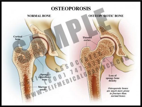
- A common condition, particularly among elderly women, osteoporosis is the reduction of bone density.
- Osteoporosis affects both the dense cortical portion on the outside of a bone and the spongy bone inside.
- Causes include age, low body mass, family history, smoking, malnutrition, alcoholism, insufficient physical activity and certain medications and toxins. It is also associated with a number of chronic diseases.
- The primary risk is that of fracture, particularly of the hip, a potentially devastating event in the elderly. In severely osteoporotic patients, healing may be slow and insufficient.
- Treatment depends on the cause but includes medication, dietary changes, and exercise.
-
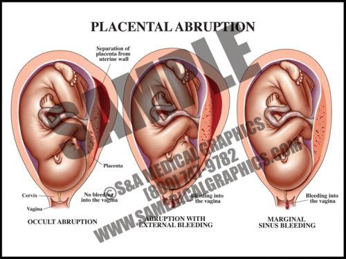
- Abruption occurs when the placenta separates from the uterine wall. Most of the time, this is accompanied by vaginal bleeding as the blood travels between the membranes and the uterus. Some- times, however, there is no bleeding because edges of the placenta remain sealed.
- Symptoms frequently include hypertonic uterus with severe abdominal pain and rapid contractions. If the abruption is large (more than about 50%), the fetus may not survive.
- The specific cause of placental abruption is often unknown, but risk factors include abdominal trauma and maternal factors like smoking, drinking, and diabetes, among others.
- Fetal distress occurs early in this condition in about half of all cases.
-
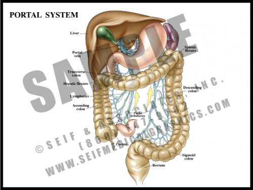
- The portal system is a specialized venous drainage system of the large bowel. Instead of merely draining deoxygenated blood, the portal system drains metabolites and nutrients upward so that they detour through the liver instead of returning directly to the heart and lungs. The liver serves as a cleaning and metabolic sieve where drugs and other chemicals are further broken down and either used or removed from the system.
-
- This 2D dissolve animation shows how this maneuver can release shoulder dystocia impaction and allow for delivery of the fetal shoulder.
- Each animation has pause and play buttons to allow for more interactivity during viewing.
-
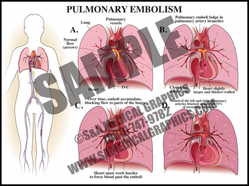
- A pulmonary embolism is a blockage in one or more arteries of the lungs. In most cases, it is caused by clots that travel to the lungs from another part of the body, most commonly from a DVT in a lower extremity.
- Depending on the size of the embolus, it can occlude the main pulmonary artery, straddle the arterial bifurcation, or dis- seminate out into the smaller branching arteries of the lungs.
- Saddle embolisms are frequently fatal, while embolic showers can be clinically silent unless they block enough of the pulmonary vasculature.
-
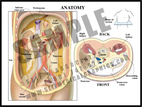
- Most of the abdominal contents lie within the peritoneum, a sac made up of a sheet of dense connective tissue. Some structures lie behind the peritoneum (retroperitoneal). Others go in and out of it, although edges are sealed and there is little or no direct connection between the intra- and retroperitoneal regions.
- The liver has a “bare area” at its top where it lies directly against the lower surface of the diaphragm, but the rest of it is intraperitoneal. The ascending and descending colons are both retroperitoneal, while the transverse colon and part of the sigmoid are intraperitoneal; the duodenum, or first portion of the small intestine, is retroperitoneal.
- True retroperitoneal structures include the pancreas, the kidneys, ureters and adrenals, the great vessels and the pelvic structures.
-
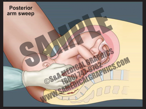
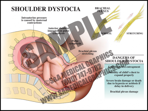
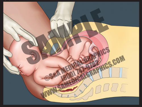
- In cases of shoulder dystocia, the upper fetal shoulder is impacted on the pubic symphysis, or more rarely, the lower shoulder becomes impacted on the sacral promontory or nonexible coccyx. In either case, this event prevents delivery of the baby. Shoulder dystocia can be a potentially catastrophic event since the fetal thorax is still within the pelvis and cannot properly expand for breathing.
- While the rate of shoulder dystocia is higher with gestational diabetic women and macrosomic fetuses (>4500 grams at birth), most cases of shoulder dystocia occur with average-sized fetuses.
- If a brachial plexus palsy (brachioplexopathy) occurs, it usually affects the portions of the brachial plexus that control the shoulder and elbow. Spontaneous recovery is the rule rather than the exception.
- Most fetuses are successfully delivered with a combination of McRoberts maneuver and suprapubic pressure.
-
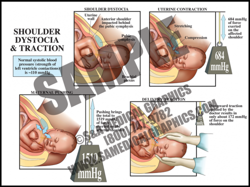
- The uterus itself generates tremendous pressure during contractions. If the average systolic blood pressure (pressure generated by the left ventricle during a contraction) is 120 mmHg, the uterus alone generates more than 5 times that amount. When the accessory muscles (the diaphragm and abdominal muscles) are used to push in conjunction with contractions, this pressure increases to more than 10 times that amount.
- The pull generated by a physician during downward traction for shoulder dystocia is slightly higher than the average systolic blood pressure and contributes only a very small amount to the total amount of generated pressures.
- Uterine and abdominal pressures are good evidence that if shoulder dystocia is the cause of brachial plexus palsy, it is most likely from the intrinsic pressures of the uterus and body wall, not the caregiver.
-
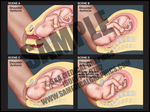
- This 2D animation demonstrates how shoulder dystocia occurs during delivery.
- It shows fetal descent down the birth canal and delivery of the fetal head, but impaction of the anterior shoulder on the pubic symphysis, preventing delivery of the shoulder.
- This animation has pause and play buttons to allow for more interactivity during viewing.
-
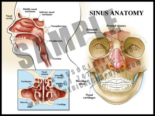
- Sinuses are hollow spaces within the facial bones. They are lined with a ciliated mucosa which has mucus glands. The sinuses are interconnected via a series of openings, allowing mucus to drain into the nose and pharynx.
- The sinuses help to warm inhaled air before it enters the lungs.
- Sinuses are prone to infection or reaction to allergens and react by mucosal swelling and overproduction of mucus. Chronic inflammation or infection can result in permanent thickening of the mucosa and reactive bone changes. Surgery is designed to facilitate drainage and relieve pressure; in some patients it must be repeated a large number of times.
-
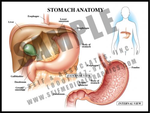
- The stomach is a muscular sac derived from the simple fetal gastrointestinal tube. The mucosal lining has specialized cells which secrete strong acids and enzymes to break food down before it passes to the small bowel for absorption and distribution.
- The walls are folded into rugae which increase the surface area of the sac. The muscular walls contract to help break up food material.
- The greater omentum arises from the greater curvature of the stomach, and the lesser omentum from the lesser curvature; the hepatoduodenal ligament lies at the free edge and contains the extrahepatic biliary ducts.
- The stomach lies under the diaphragm and to the left of the liver. The strong pyloric sphincter divides the distal stomach from the duodenum or the first portion of the small intestine.
-
- This 2D dissolve animation shows how this maneuver can release shoulder dystocia impaction and allow for delivery of the fetal shoulder.
- Each animation has pause and play buttons to allow for more interactivity during viewing.
-
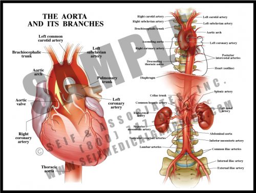
- The largest artery in the body, the aorta leaves the left ventricle and supplies all of the body’s tissues, including the heart itself, via its branches.
- The major parts are the arch (from the aortic valve to the left subclavian artery), descending thoracic aorta (from the left subclavian to the diaphragm) and abdominal aorta (from the diaphragm to the iliac bifurcation at about the level of the umbilicus).
- There are three major vessels leaving the arch and these supply the head, neck and arms; there are segmental vessels supplying the body wall throughout the length of the aorta. Major branches supply abdominal structures; the iliac arteries and their branches supply the pelvis and legs.
-
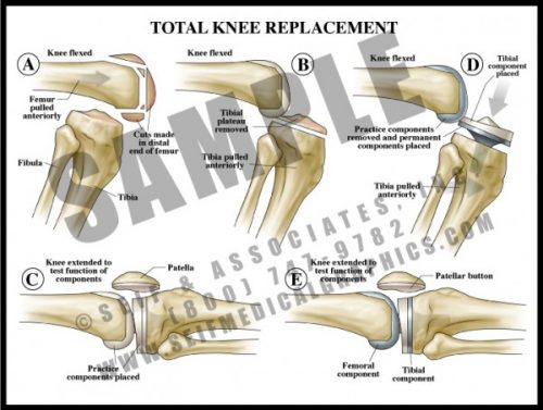
- One of the more common surgical procedures, diseased bone and articular surfaces (usually from either trauma and/or degenerative joint disease) are surgically removed.
- Most knee prostheses are made of a strong metal with a durable plastic overlay to serve as a joint surface.
- As with any bony prosthesis, these can be either cemented into place or have a roughened surface which allows bone to grow into the surface.
- All three units (femoral, tibial and patellar) can be inserted together or individually, depending upon the specific needs of the patient.