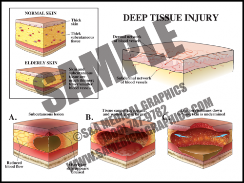
- Deep tissue injuries can have the same outward appearance as decubitus ulcers, but the underlying etiology is different. These injuries happen and progress quickly.
- The mechanism of this injury is pressure to the skin and soft tissue, within a short period of time, that compromises tissue perfusion and results in ischemia and damage to the deeper subcutaneous tissues.
- The initial injury is not visible on the surface of the skin and only manifests later as the underlying tissue starts to necrose, first forming what appears to be a bruise before progressing to an external/visible skin wound.
- This type of wound evolves upward towards the skin as well as deeper, so once the external wound becomes apparent, it is frequently already a deep injury often with the appearance of a Stage 3–4 decubitus ulcer.
-
-
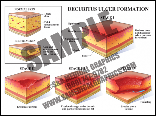
- Pressure ulcers are a localized injury to the skin and/or underlying tissue, usually over a bony prominence, that occur as a result of pressure, shear, and/or friction.
- Stage I decubitus ulcers manifest as a non-blanchable area of skin redness, which then progresses to a partial thickness wound in Stage II, and further to full thickness skin loss in Stage III. Stage IV decubiti involve full thickness tissue loss with exposed bone, tendon, or muscle with sloughing, undermining, and tunneling. These ulcers can extend into muscle and/or supporting structures making osteomyelitis and sepsis a concern.
- Decubitus ulcers are associated with an increased morbidity and mortality, and healing can be difficult as debilitated patients who form them usually have widespread vascular disease, nutritional de cits, and/or oxygenation/perfusion difficulties.
-
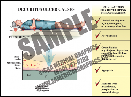
- Decubitus ulcers are common in debilitated patients, particularly the aged who have thinner skin and subcutaneous tissue that is more susceptible to compression injury.
- Patients with limited mobility (from injury, sickness, or neurologic disorders, etc.), poor nutritional intake, conditions affecting perfusion and oxygenation of tissues (like diabetes, cardiovascular disease, heart failure, etc.), skin moisture due to incontinence, and advanced age are particularly vulnerable.
- These ulcers are often multifactorial, making them difficult to prevent and even more difficult to heal in at-risk patients.
-
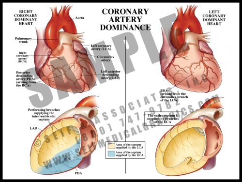
- Right heart dominance: the posterior portion of the interventricular septum is supplied by the posterior descending branch of the right coronary artery.
- Left heart dominance: the entire septum is supplied by branches of the left anterior descending artery; an obstruction in that vessel may lead to loss of the entire septum, an often fatal event. The posterior descending artery is derived from a branch of the circumflex artery instead of from the RCA.
-
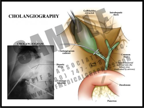
- Performed during surgery for gallbladder removal, this is an effective intraoperative radiographic test to look for either blockage or leakage in the biliary tree.
- This test may be performed prior to removing the gallbladder, or at any time a problem is suspected. A tiny catheter is threaded through a small incision in the cystic duct. Dye is injected into the biliary tract and x-rays are taken, allowing the surgeon to see which ducts are patent. Voids represent stones or tumors, and extravasation represents a leak in the system.
- While this test is very reliable in the case of a retained stone or suspected damage, the outcome in patients having routine intraoperative cholangiography without apparent complication is the same as those in whom the test was not performed.
-
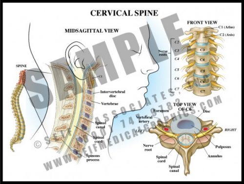
- The 7 vertebrae of the cervical spine help support the skull and protect the spinal cord as it exits the cranium to pass downward through the spinal canal.
- The transverse processes each have a small hole through which the vertebral arteries pass to join to form the basilar artery supplying the posterior and deep portions of the brain and brainstem.
- The cervical nerve roots form the brachial plexus which supplies sensation and movement to the upper extremities.
- Degenerative joint disease and disc disease are very common in the cervical spine, leading to arm and hand pain and dysfunction requiring decompression and sometimes fusion.
-
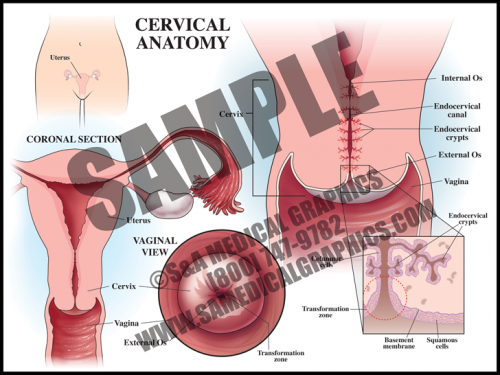
- The cervix is the circular muscle at the base of the uterus; it makes up the top of the vagina.
- The cervical canal passes through the cervix and allows blood from menstruation and a fetus to pass from the uterus into the vagina.
- During a pap smear, a screening test for cervical cancer, cells are scraped from the opening of the cervix and examined under a microscope for abnormality.
- The transformation zone is the area where the mucus secreting columnar cells of the endocervix meet the squamous cells of the ectocervix. Most squamous cell carcinomas and dysplasias are found here.
-
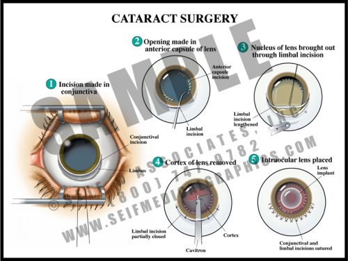
- The lens opacifies with age or after trauma. When vision is sufficiently affected, the cataract can be surgically removed and replaced with an artificial intraocular lens.
- The cornea is lifted from an incision in the blue-grey line surrounding the iris, and the anterior surface of the lens is opened. The nucleus of the lens is removed, leaving the posterior capsule of the lens in position.
- The intraocular lens is then placed within the capsule and fixed into position. Laser treatments are often needed post-operatively to clear the posterior capsule.
- This procedure is one of the safest and most common surgical procedures performed today, with a very low rate of complications.
-
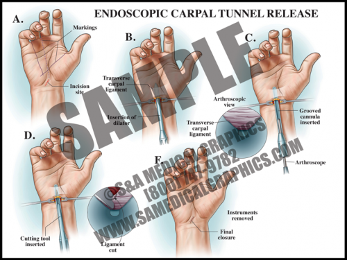
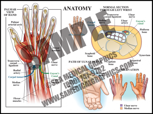
- If conservative treatment is not effective, surgical intervention may be chosen. Surgery can be done as an open procedure or endoscopically.
- In endoscopic surgery, a small incision is made in the wrist and a dilator tube is inserted. An arthroscope with a camera is inserted in order to view the anatomy, then a cutting tool is used to sever the transverse carpal ligament in order to reduce pressure on the median nerve.
- This type of surgery is less invasive and allows for faster recovery and less postoperative pain.
-
- The carpal tunnel is a passageway on the palmar side of the wrist that houses the median nerve and finger flexor tendons. Anteriorly, the carpal tunnel is bordered by the transverse carpal ligament, a heavy band of fibers that forms the fibrous sheath containing the carpal tunnel; posteriorly, the carpal tunnel is bordered by carpal bones.
- Guyon’s canal contains the ulnar nerve and artery; this anatomy does not pass through the tunnel, but lies superficial to it.
- Carpal tunnel syndrome occurs when the median nerve, which controls sensations to the palm side of the thumb, first, second, and half of the third finger, becomes compressed at the wrist, within the carpal tunnel. This results in pain, weakness, or numbness in the hand and wrist, radiating up to the arm.
-
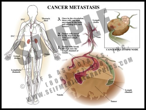
- Cancer tends to spread by two mechanisms: infiltration, in which the tumor pushes against and enters contiguous tissue; and metastasis, when cancer cells enter lymphatic channels and/or small blood vessels and eventually travel to distant locations and organs in the body.
- Certain tumors have a predilection for specific sites. Colon cancer frequently spreads to the liver, and breast cancer to the brain and spine.
- When there is lymphatic spread, the local and regional lymph nodes are the first line of defense; when the nodes fill up with dividing tumor cells, the cells then break free and travel toward the heart for distribution throughout the body.
-
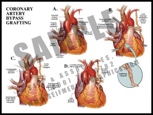
- When coronary arteries are significantly blocked (>70% stenosis), pain symptoms often occur with exercise in the form of stable angina, or at rest in the form of unstable angina.
- Bypass vessels are harvested as free grafts from the saphenous veins of the legs. These are anastomosed to the aorta and then to the coronary arteries, literally bypassing the blocked regions.
- The internal mammary arteries, which lie on either side of the sternum within the rib cage, can also be harvested and anastomosed directly to the coronary arteries. These grafts are less likely to stenose than are vein grafts.
- The procedure can be performed either on or off cardiopulmonary bypass.
-
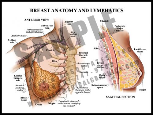
- The breast in a premenopausal woman is composed of glandular tissue, fat, connective tissue and ducts; the axillary tail of breast tissue is tucked upward in the axilla. The breast lies on the pectoralis muscles of the thorax.
- The breast is divided by irregular fibrous septa which prevent masses from migrating from one area of the breast to another; malignant tumors may eventually erode through these septa.
- Lymphatic channels travel throughout the breast, with all but the most medial portions draining to the axillary lymph nodes. The “sentinel” node—the first node to receive drainage from the breast—can be determined with testing and evaluated for metastatic spread.
- Post-menopausal women have little glandular tissue since most of it has been replaced by fat.
-
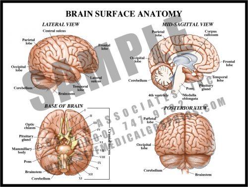
- The surface of the brain has multiple folds, or gyri, separated by sulci. While many of these are specific to a particular individual, some are constant and serve as landmarks for functional control.
- The cerebrum is the large, rounded portion of the brain and is the site of higher functions. The cerebellum and brainstem control more basic functions like heart rate, balance, and respiration.
- The cranial nerves mostly originate in the brainstem and exit the skull via foramina in the base of the skull (see M1). The optic nerves originate in the occipital lobes, travel along pathways inside the brain matter, and exit anteriorly.
- The corpus callosum is a group of myelin-covered neuronal fibers (white matter) which connect the right side of the brain with the left.
-
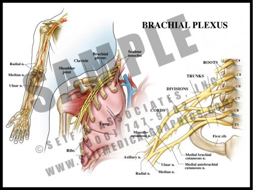
- The nerve roots of the lower cervical spinal cord split and merge several times before supplying the arm and hand.
- The brachial plexus lies over the first rib and behind the clavicle. It is intimately related to the subclavian/brachial artery and passes between the scalene muscles of the neck.
- The plexus is divided into roots, trunks, divisions, cords and terminal branches. By looking at the anatomical distribution of pain or dysfunction, it is possible to determine the location of a brachial plexus lesion.
- Brachial plexopathy can occur during delivery with or without shoulder dystocia, and from thoracic outlet syndrome.
-
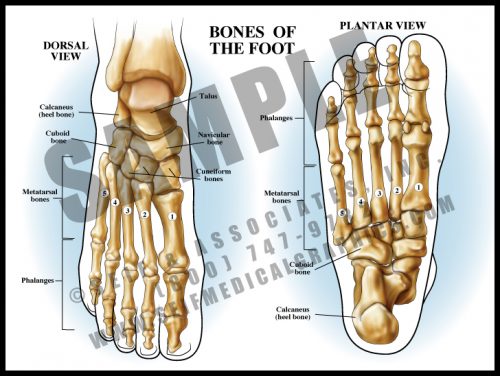
- The bones of the foot follow basically the same pattern of the hand bones. There is a double layer of sesamoid-like bones forming the ankle, which articulate with the long bones making up the central foot (metatarsals), which are in turn attached to the phalanges, or toes.
- The tendons and the many ligaments of the foot attach to the tough, thin tissue covering the bones, the periosteum. The ligaments attach the bones to each other, and the tendons connect the muscles to the bones.
- As in the hand, there are intrinsic muscles in the feet.
- The bones of the foot form a longitudinal arch and a transverse arch. Most of the body’s weight is borne on the metatarsal heads, particularly the first and fifth.
-
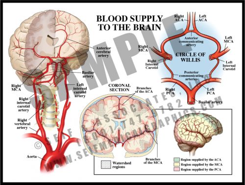
- The anterior 2/3 of the brain is supplied by branches of the internal carotid artery, whose terminal branches form the anterior and middle cerebral arteries.
- The vertebral arteries branch off the subclavian arteries and through small openings in the transverse processes of the cervical vertebrae. They merge to form the basilar artery supplying the cerebellum, brain stem, and the posterior cerebrum via the posterior cerebral arteries.
- The Circle of Willis has small connecting vessels between the three major cerebral vessels. Blood can change direction within the circle for collateral blood flow if needed. There can be significant variation in the form of the Circle of Willis from one individual to another.
- Tissues supplied by the tiny terminal branches of vessels are known as watershed regions and are vulnerable to damage during periods of low perfusion or oxygenation.
-
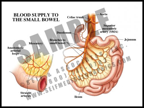
- With the exception of a portion of the first part of the duodenum, the small bowel is supplied by the many branches of the superior mesenteric artery.
- The branches anastomose with each other in two layers of arcades or arches, and from these, small straight vessels pass to the bowel surface, traveling around and through the wall, dividing into smaller and smaller branches.
- The arcades and multiple straight vessels are an adaptation which protects the bowel. Damage can occur to a portion of the small bowel without loss of the entire organ. Clots and ischemia from atherosclerosis and other vascular pathologies can affect the small bowel, much like the brain, heart, kidney and other organs can be affected by such conditions.
-
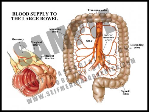
- The blood supply of the colon comes from three sources: the superior mesenteric arteries supplying the cecum, ascending (right) colon and half of the transverse colon; the inferior mesenteric arteries supplying the distal half of the transverse colon, the descending (left) colon, and the sigmoid colon; the rectal arteries supply the rectum.
- The arteries then divide into arcades, as they do to the small bowel, with straight arteries entering the bowel wall at the mesenteric border.
-
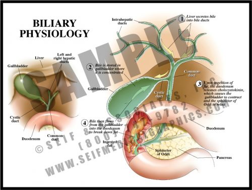
- The gallbladder stores bile formed within the liver, releasing it for fat digestion.
- Bile travels through the intrahepatic ducts into the paired hepatic ducts; these merge into the common hepatic duct. Bile is then diverted via the cystic duct to the gallbladder for storage.
- When food is ingested and travels through the stomach to the duodenum, a hormone is released (cholecystokinin) which stimulates the gallbladder to contract and the sphincter of Oddi to relax. This allows bile to flow through the cystic duct and the common bile duct into the duodenum.
- The most common pathology in the extrahepatic biliary system is bile (gall) stones (concretions of bile salts, cholesterol, and minerals) which can block ducts, causing inflammation, pain, and jaundice.
-
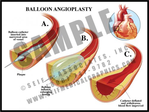
- This procedure is a relatively non-invasive technique of opening stenotic blood vessels.
- A catheter is threaded through the arterial system from the arm or leg and into the diseased artery. The balloon is then positioned inside the stenotic area and gently inflated several times to crush the plaque and flatten it against the walls of the vessel.
- This procedure is commonly performed and is often accompanied by the deployment of a stent to hold the vessel open.
- Complications can include clot formation on the fractured plaque after the release of clotting factors and formation of a dissection (sometimes incorrectly called a “dissecting aneurysm”) in the vessel wall.
-
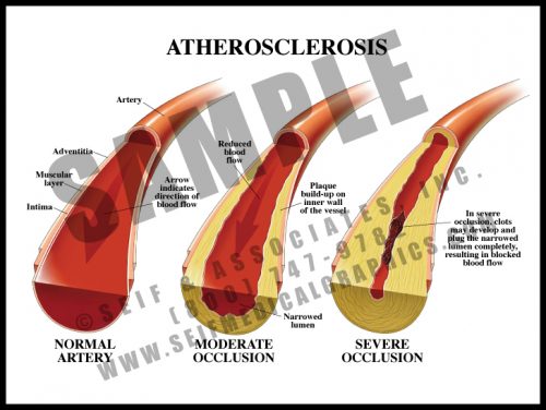
- Atherosclerotic plaque is fatty, cholesterol-laden material which accumulates within the inner layer of the major arteries, narrowing the diameter of the lumen or opening.
- It can occur in any artery in the body and is a direct cause of stroke when in the carotid arteries; myocardial infarction when in the coronary arteries; acute bowel ischemia when in the mesenteric vessels; peripheral vascular disease when in vessels to the legs, etc.
- Atherosclerosis can result in increased blood pressure in an effort to overcome the higher pressures caused by arterial stenosis throughout the body.
-
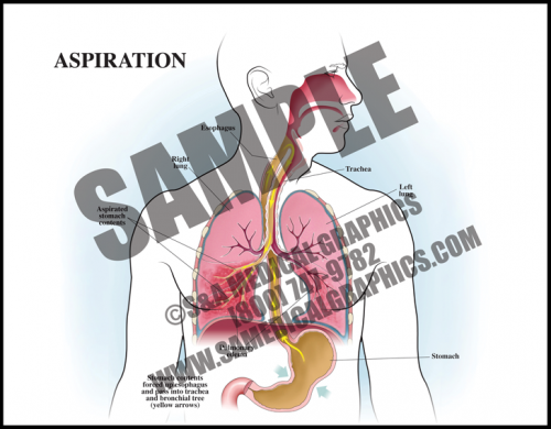
- Aspiration occurs when foreign material, of either oropharyngeal or gastric contents, is inhaled into the lungs.
- Aspiration can cause a number of respiratory problems depending on the quantity and nature of the inhaled material. Aspiration of gastric contents causes pulmonary edema and often pneumonia.
- The risk of aspiration is increased by conditions associated with altered or reduced consciousness, esophageal conditions like dysphasia, certain neurological disorders, and mechanical conditions like NG tube placement, endotracheal intubation, etc.
-
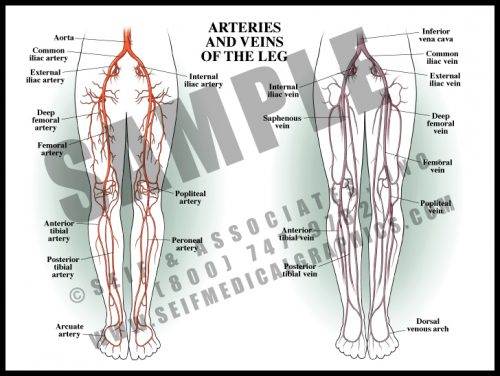
- Like in the arm, the deep, muscular arteries of the leg travel together, but the superficial veins are unpaired and variable in course.
- The legs receive blood from the terminal branches of the aorta, the iliac arteries. Branches supply the muscles of the thigh, and the name of the artery changes as it passes certain landmarks.
- The major vessels trifurcate behind the knee, dividing into the anterior and posterior tibial arteries and the peroneal artery, all of which travel toward the feet supplying the muscles and other tissues along the way.
- As in the arm, there are connective vascular arches in the foot supplying collateral circulation.
-
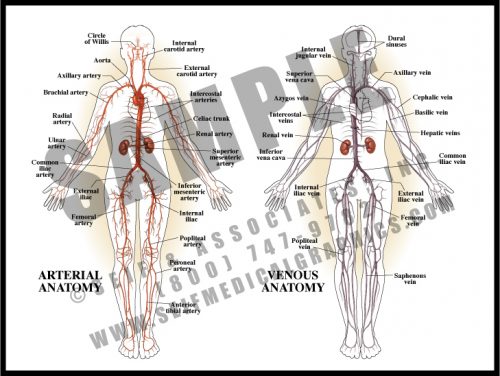
- Although the cardiovascular system is referred to as one unit, it is actually two separate systems which work independently.
- Through the arterial supply, oxygenated blood is distributed from the lungs to the left heart and aorta, and eventually to within 5 cells of every cell in the body. The arteries divide into smaller arteries, then into arterioles, which in turn divide into capillaries. Oxygen exchange takes place at the level of the capillaries, vessels whose walls are only one cell thick.
- In the venous system, deoxygenated blood drains from the capillaries, which conjoin into venules, small veins, veins, and the major draining vessels – the superior and inferior venae cavae. This blood then enters the right heart and travels to the lungs to re- oxygenate and start the cycle again.
-
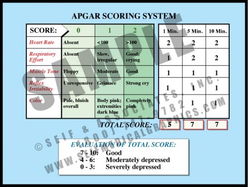
- This simple scoring system indicates how well the fetus fared through labor and delivery. The maximum score is 10 and the minimum is 0, with up to 2 points being awarded for each of 5 measures of health.
- Apgar scoring is done at 1 minute and 5 minutes of age, sometimes by nursing staff, sometimes by physicians. If the 5-minute score is low, a 10, 15 or 20-minute score may be recorded, either until the baby is stable or moved from the delivery room for specialized care.
- While there is some correlation between low 5 minute Apgar scores and neurological outcome, only a very small percentage of children with low 5 minute scores sustain brain damage. The longer the score remains low, the higher the correlation with neurological damage.
-
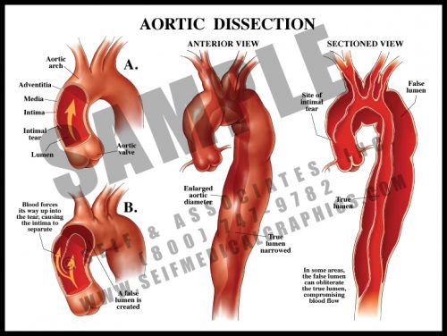
- Sometimes called “dissecting aneurysm”, this is not an aneurysm, but a separation of the aortic wall layers.
- Blood enters the aortic wall through a small tear in the intima or inner lining of the artery. Under pressure, it then dissects through the wall, creating a false lumen or false channel. Sometimes there is a second tear through which the blood re-enters the true aortic lumen; sometimes the blood breaks through the wall to the thorax or retroperitoneal spaces.
- Dissections are usually associated with hypertension and atherosclerosis, although certain genetic conditions (Marfan’s syndrome) can predispose to dissection.
- Symptoms include a severe tearing pain in the back as the dissection travels distally, changes in blood pressure and distal pulses, and loss of various physiologic functions if the dissection blocks the blood supply to major organs.
-
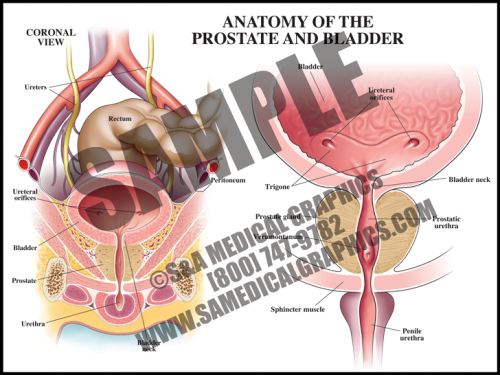
- The prostate is a walnut-sized gland located between the bladder and the penis; it secretes fluid that nourishes and protects sperm. The male urethra runs through the center of the prostate, from bladder to penis.
- The bladder is a hollow, muscular organ in the lower abdomen that stores urine and allows urination to be infrequent and voluntary.
- It is not uncommon for older men to develop benign prostatic hyperplasia (BPH), in which the prostate becomes enlarged, resulting in restriction of the ow of urine through the urethra. The prostate can also develop cancer, although that is much less common than BPH.
-
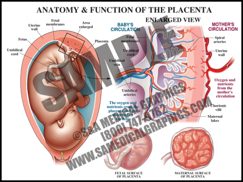
- The placental is a flattened, circular organ that develops in the uterus during pregnancy in order to support the fetus. The fetus is attached to the placenta via the umbilical cord.
- The placenta transfers oxygen and nutrients from the maternal blood to the fetal blood; it also transfers wastes products from the fetal blood to the maternal blood for removal.
- The umbilical cord is normally comprised of three blood vessels, two smaller umbilical arteries that carry deoxygenated blood from the fetus to the placenta, and a larger umbilical vein that supplies the fetus with oxygenated blood from the mother.
- The placenta is expelled from the uterus after the fetus is delivered. Placental pathology can reveal important information about intrauterine conditions and events, as well as shed light on the cause of adverse fetal outcome.
-
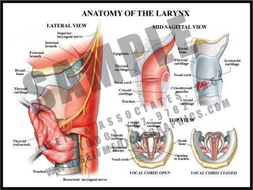
- The larynx is composed of a number of cartilaginous structures, muscles and ligaments which maintain the patency of the airway and hold the vocal cords under tension during speech.
- The large thyroid cartilage, which lies beneath the thyroid gland, is connected to the hyoid bone by a strong ligament (thyrohyoid ligament), and the epiglottis arises from its internal surface. All internal structures with the exception of the vocal cords are covered by a pink mucosal lining.
- The small cartilages to which the vocal cords are attached are moved by tiny muscles under the control of the recurrent, superior and inferior laryngeal nerves. These muscles make small adjustments in the opening between the cords, allowing different pitches of sound to be created.