34-year-old male was involved in an MVA in which he suffered significant cervical spine fractures. He presented to the defendant institution for evaluation and treatment. An x-ray, CT, and MRI were all done that evening which showed fractures of the C6 and C7 facets bilaterally, a burst fracture at C7, laminar fractures at C7, and subluxation of C6 over C7 with perched facets. The surgeon who reviewed the films that night noted no disk herniation and only cord contusion. He failed to mention the subluxation at C6-7 and went home for the night.
The patient was placed in a Miami J-collar and observed that evening. The nursing staff assigned to the patient gently rolled him on occasion to perform standard tasks. Early the next morning, the patient was noted to have lost all movement and feeling from the chest down. The repair surgery was ultimately done to stabilize the spine, but the patient never completely recovered neurologically and remains a paraplegic.
The plaintiff claims the nurses never should have rolled the patient given his condition, which resulted in his paraplegia. The defense (the hospital and the nursing staff) counters by saying the surgeon who reviewed the films never made mention of the instability of the patient’s cervical spine to the nursing staff and never gave any orders that the patient should not be rolled. Given the instability of the fracture based on the MRI evidence, the surgeon should have taken the patient in for emergency surgery immediately after reviewing the films.
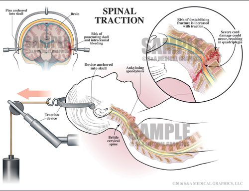
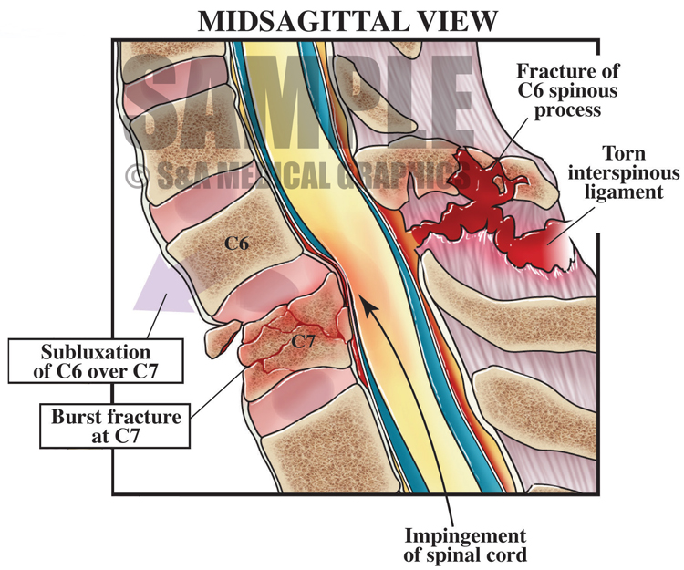
The first exhibit was intended to show the nature of the patient’s cervical spine fractures and to show the relative instability of the C6–C7 subluxation and bilateral facet fractures.
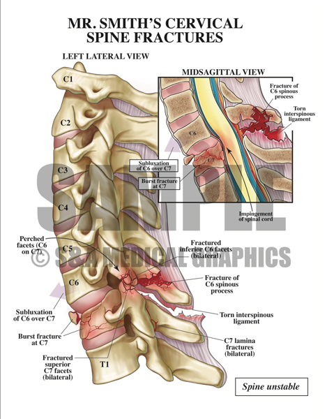
Exhibit 1: Illustration depicting the nature of the patient’s cervical spine fractures.
The next exhibit was a series of CT images that best demonstrated the cervical fractures along with an overlay showing full-color artistic interpretations of these film images.
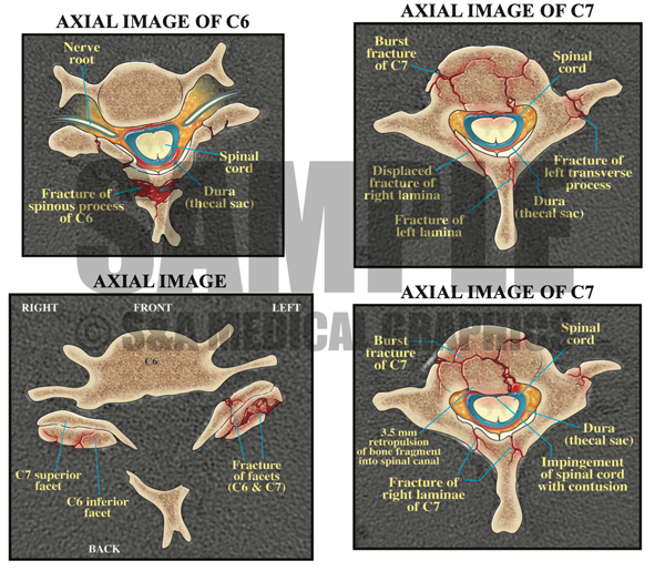
Exhibit 2: CT images with illustrated overlays that best demonstrate the fractures in this patient.
Another exhibit showed the MRI films and interpretive illustrations.
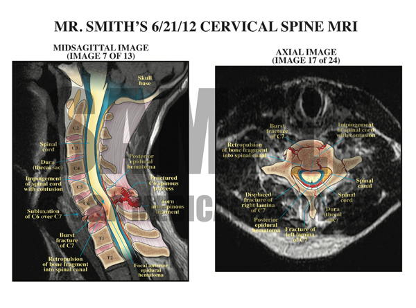
Exhibit 3: Cervical spine MRI interpretations showing the various fractures and associated soft tissue damage.
The last exhibit is yet another CT film interpretation of the cervical fractures focused on the bilateral facet fractures.
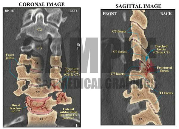
Exhibit 4: CT interpretations focusing on the bilateral facet fractures.
During testimony with all of these fracture exhibits, it was made clear to the jury that the instability of the cervical spine was NOT made known to the nursing staff at the defendant institution.
—Editorial & illustrations contributed by Russ Edwards, MS, CMI