This case involved a female patient with a congenital dental condition in which she was missing teeth #7, #10, and #11 (total of 10 teeth) who presented to the defendant oral surgeon for placement of dental implants and crowns. On 3/19/12, Dr. Jones placed the dental implants – one at #7 and one at #11 – using bone graft to augment the alveolar bone to better support the implants. The implant placement was excellent, and the final restorative dental work (abutments and crowns) greatly improved the patient’s smile and overall appearance.
The patient did well for several years until January of 2019 when she began to note tooth pain, difficulty chewing, and loosening of the dental implants. She presented to another dentist on 1/15/19 and was found to have infected implants and impending implant failure at both #7 and #11. She was referred to another oral surgeon for removal of the implants, which was done on 1/30/19.
Plaintiff claims that the defendant failed to properly perform the implant surgery of 3/19/12, specifically by not placing enough bone graft to support the implants long-term. This resulted in implant failure and the need for revision surgery. The defense argues that the implants did well for nearly 7 years and that had the implants been improperly placed, they would have failed within the first 2 years after the surgery. This did not happen in this case. Her onset of symptoms occurred relatively quickly, suggesting that the failure was likely due to a more recent event such as grinding of teeth during sleep or gingival infection.
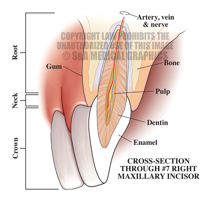
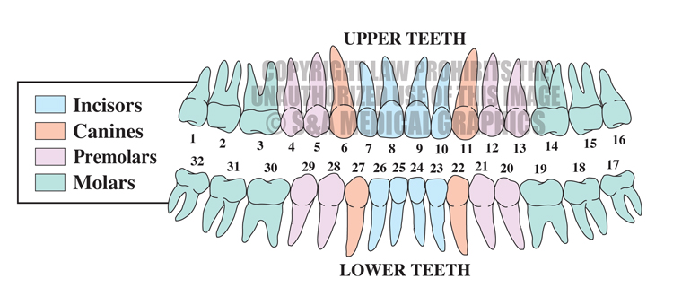
We used the first exhibit to help provide a baseline understanding of the anatomy to be discussed at trial. This also provides the numbering system for teeth, which would be critical to understanding which teeth were involved in this patient.
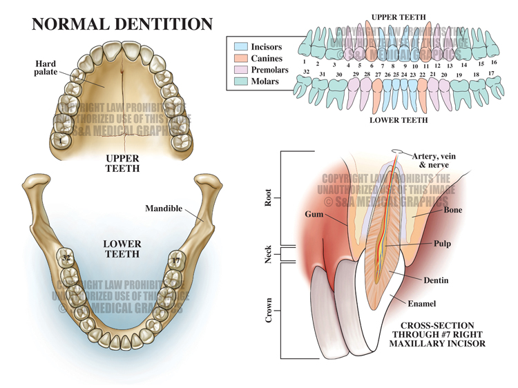
Exhibit 1 normal anatomy and numbering system for teeth.
The next exhibit detailed her dental condition prior to any restorative work, with missing teeth indicated and properly numbered.
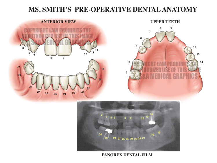
Exhibit 2 depicts the patient’s dental condition prior to surgery.
A subsequent exhibit showed the major steps involved in the implant placement surgery performed by the defendant.
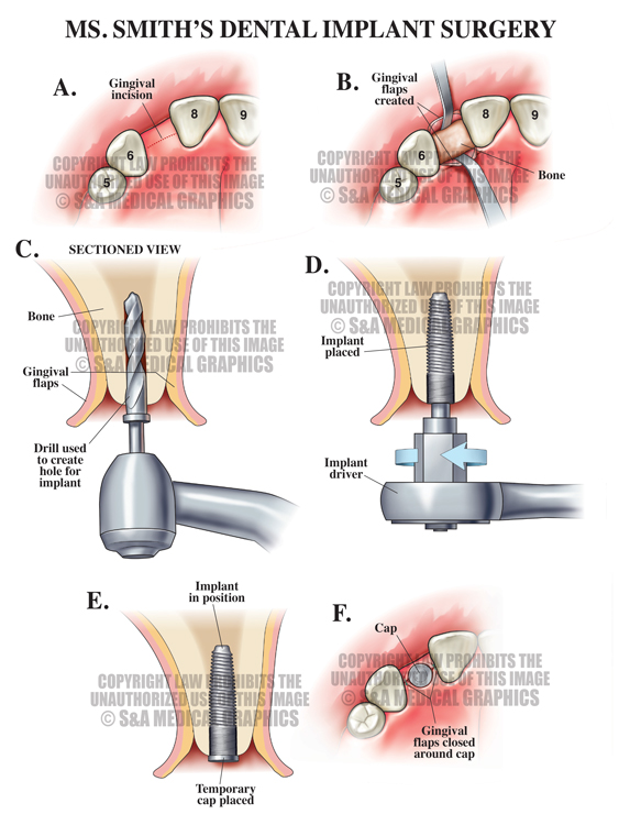
Exhibit 3 depicts the dental implant surgery steps.
An exhibit mirroring the pre-procedure condition board was created to show the much-improved appearance of her teeth after the surgery and placement of the crowns.
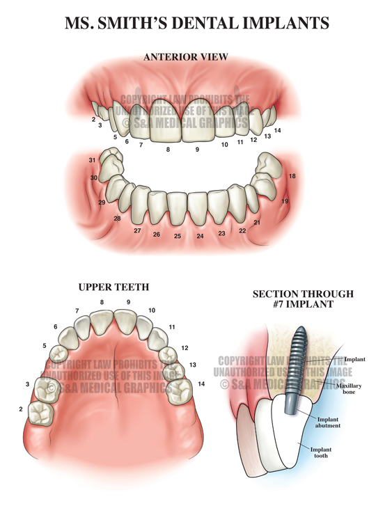
Exhibit 4 depicts the patient’s condition after the surgical procedure.
An exhibit was needed to explain the mechanisms by which dental implants can loosen through no fault of the defendant or his procedure – specifically how grinding of the teeth or periodontal infection can cause the implants to loosen over time.
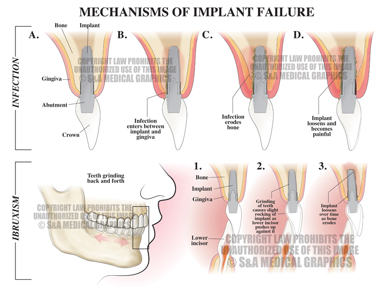
Exhibit 5 depicts how dental implants can loosen.
Finally, we developed a double-wide timeline graphic that indicated the time of the surgery at issue, the time of the first complaints of tooth pain almost 6 years later, and the times where she was seen by other dentists/oral surgeons. The focus of this exhibit was to show the expected time frame for implant failure attributable to the implant surgery relative to the span of time during which this patient had no complaints—2 years versus almost 6 years. Clearly, the implant failure was not due to the defendant’s procedure, based on this timing.
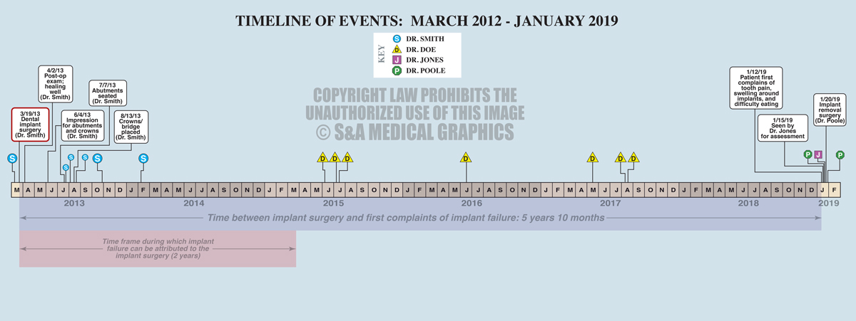
Exhibit 6 depicts the timeline of events.
—Editorial & illustrations contributed by Russ Edwards, MS, CMI