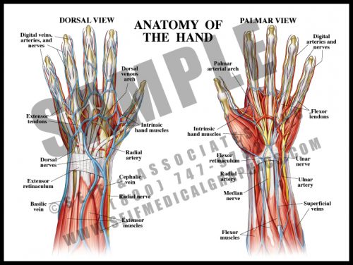
- One of the more complex structures, the hand has a high concentration of nerves and vessels. There are also many small intrinsic muscles which allow fine motor function.
- Tendons, blood vessels and nerves originating in the arm cross the wrist to enter the hand; the intrinsic muscles are solely within the hand.
- The structures crossing the double row of wrist bones are held in place by the flexor retinaculum on the volar (palmar) side (“carpal tunnel”), and by the extensor retinaculum on the dorsal side. The flexor retinaculum can sometimes thicken or scar, causing compression of the median nerve, or carpal tunnel syndrome.
-
-
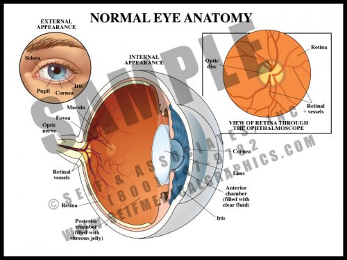
- The external eye has upper and lower lids which close over the globe to protect it. The sclera is the white of the eye, the colored portion is the iris, and the black opening in the middle of the iris is a hole known as the pupil. This is the only window in the body through which the nervous system can be seen directly.
- The anterior transparent media consists of the cornea, anterior chamber and lens; the posterior elements of the globe are covered with specialized nerve tissue, the retina.
- The optic nerve enters the eye posteriorly along with its own blood supply; this area is known as the optic disc. The macular area is where visual acuity is greatest.
-
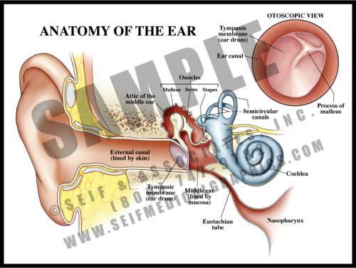
- The external ear acts as a collecting device for sound waves, focusing them into the canal.
- External sound waves cause vibration of the tympanic membrane (ear drum). The vibrating membrane moves the three ossicles of the middle ear (malleus, incus, and stapes) which transfer the vibration to branches of the vestibulocochlear nerve (cranial nerve VIII) within the cochlea.
- Motion and balance are detected by three fluid-filled canals in the temporal bone. Oriented in three perpendicular planes, the canals contain tiny hair cells that pick up fluid movement with motion of the head. This information is transmitted through the vestibular portions of the nerve to the appropriate portions of the brain.
-
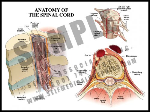
- The spinal cord lies within the spinal canal, formed by the vertebral bodies and the bony arch formed by the pedicles and laminae (see M6).
- The cord and its terminal nerves, the cauda equina, lie within the dural sac, a tough membranous structure filled with cerebrospinal fluid bathing and protecting the cord.
- The spinal cord itself ends at the level of the first lumbar vertebra, but nerve roots travel inside the dural sac to exit at lower levels; these roots form the cauda equina (”horse’s tail”).
- Blood supply comes from the segmental branches of the aorta, traveling along the nerve root to emerge as the anterior spinal artery running in the front midline of the cord; there are two parallel vessels along the back surface of the cord. There is also a generous venous plexus within the canal.
-
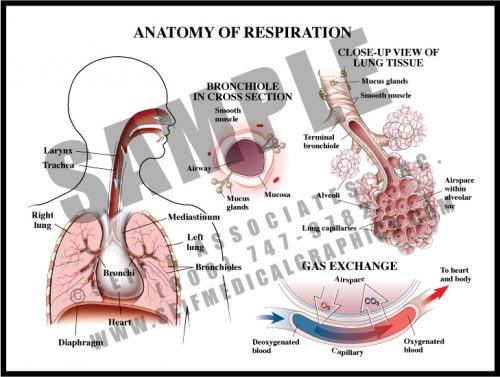
- The lungs are composed of thin-walled alveoli whose sacs are covered by a meshwork of capillaries. This is where oxygen and carbon dioxide are exchanged.
- The trachea carries air from the nose and mouth to the bronchi, which branch to each lung. These divide several times to become very small bronchioles, which directly supply the alveoli.
- The airways are lined with a ciliated mucosa which carries debris upward to the mouth on a layer of mucous, where it is swallowed. These mucosal membranes can swell in reaction to allergens, bacteria and viruses, leading to narrow airways and respiratory symptoms.
-
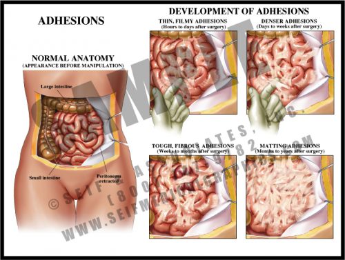
- Adhesions are fibrous scars which can form after any disturbance within body cavities and spaces. Inciting events can include surgery, trauma, and inflammation.
- Within hours of disturbance, thin, filmy strands form between bowel loops and between bowel and peritoneum and/or body wall. These continue to form for a period of time and mature over a period of weeks.
- Mature adhesions are dense, white fibrous tissues which have merged with the outer layer of the tissues; they eventually develop their own blood supply and may become severe enough to cause chronic pain and pose a chronic risk of small bowel obstruction or volvulus.
-
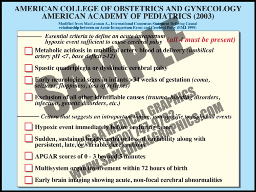
- In 2003, the American College of Obstetrics and Gynecology and the American Academy of Pediatrics recommended adoption of a definition of term intrapartum asphyxia developed by an international task force.
- The definition requires that all four parameters must be met in order to diagnose intrapartum asphyxiation in a term fetus. These include metabolic acidosis, diagnosis of specific types of cerebral palsy correlated with the types of asphyxia damage seen in term fetal brains, early seizures and other neurological signs, and exclusion of all other causes.
- There are other criteria that suggest an intrapartum timing of injury, nonspecific to asphyxia event. These include a hypoxic event immediately before or during birth, sudden, sustained bradycardia or loss of variability along with persistent, late, or variable decelerations, low APGAR scores (<3) beyond 5 minutes, multisystem organ involvement within 72 hours of birth, and early brain imaging showing acute, non-focal cerebral abnormalities.
-
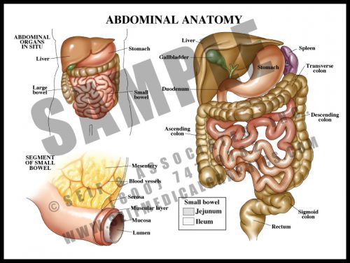
- The contents of the abdomen are primarily associated with digestion and distribution of nutrients.
- The esophagus, a tube which carries food and fluid through the thorax, enters the abdomen through the diaphragm, where it widens into the stomach; the stomach empties into the small bowel (duodenum, jejunum, and ileum, in which food is absorbed into the blood stream), and from there into the large bowel, where waste material is compacted as fluid is reabsorbed into the system.
- The liver has multiple functions affecting a number of other body systems, including digestive, hematologic and endocrine/metabolic.
- The large and small bowels are supplied by branches off of the aorta carried within the mesentery, a double-layered sheetlike structure.