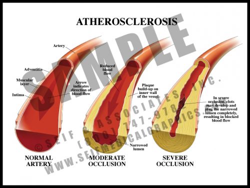
- Atherosclerotic plaque is fatty, cholesterol-laden material which accumulates within the inner layer of the major arteries, narrowing the diameter of the lumen or opening.
- It can occur in any artery in the body and is a direct cause of stroke when in the carotid arteries; myocardial infarction when in the coronary arteries; acute bowel ischemia when in the mesenteric vessels; peripheral vascular disease when in vessels to the legs, etc.
- Atherosclerosis can result in increased blood pressure in an effort to overcome the higher pressures caused by arterial stenosis throughout the body.
-
-
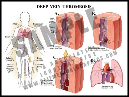
- These potentially lethal blood clots usually form in the deep veins of the leg or in the pelvis. Those in the legs are usually painful, whereas those in the pelvis may be asymptomatic.
- The clots usually form in the valves of the larger veins, propagating upward toward the heart.
- Clots of any size can break off and travel with the blood flow through the inferior vena cava to the right side of the heart and to the lungs; the pulmonary vasculature acts like a sieve and clots get caught in the vessels as the vessels get smaller, causing loss of blood flow in those areas. These clots are known as pulmonary emboli if they reach the lungs.
- Conditions associated with DVT and PE include a history of leg trauma, cancer, surgery, venous stasis from illness, lack of exercise, clotting defects and others.
-
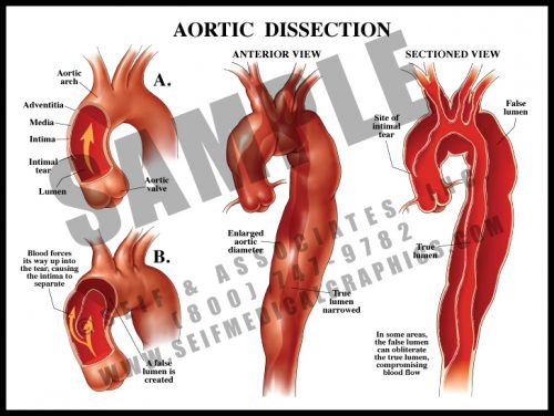
- Sometimes called “dissecting aneurysm”, this is not an aneurysm, but a separation of the aortic wall layers.
- Blood enters the aortic wall through a small tear in the intima or inner lining of the artery. Under pressure, it then dissects through the wall, creating a false lumen or false channel. Sometimes there is a second tear through which the blood re-enters the true aortic lumen; sometimes the blood breaks through the wall to the thorax or retroperitoneal spaces.
- Dissections are usually associated with hypertension and atherosclerosis, although certain genetic conditions (Marfan’s syndrome) can predispose to dissection.
- Symptoms include a severe tearing pain in the back as the dissection travels distally, changes in blood pressure and distal pulses, and loss of various physiologic functions if the dissection blocks the blood supply to major organs.
-
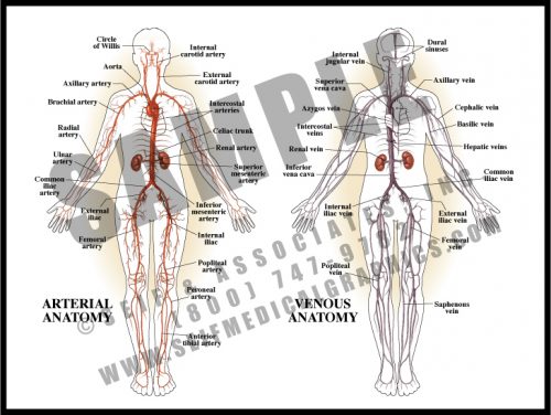
- Although the cardiovascular system is referred to as one unit, it is actually two separate systems which work independently.
- Through the arterial supply, oxygenated blood is distributed from the lungs to the left heart and aorta, and eventually to within 5 cells of every cell in the body. The arteries divide into smaller arteries, then into arterioles, which in turn divide into capillaries. Oxygen exchange takes place at the level of the capillaries, vessels whose walls are only one cell thick.
- In the venous system, deoxygenated blood drains from the capillaries, which conjoin into venules, small veins, veins, and the major draining vessels – the superior and inferior venae cavae. This blood then enters the right heart and travels to the lungs to re- oxygenate and start the cycle again.
-
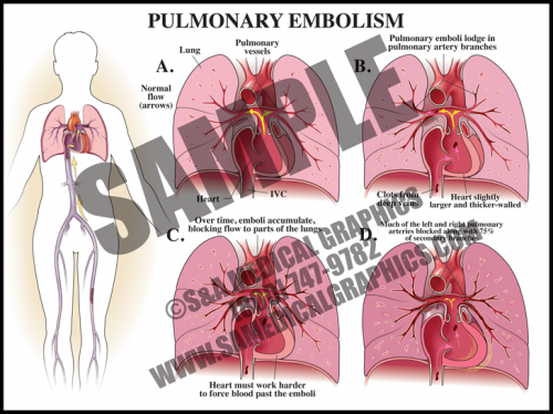
- A pulmonary embolism is a blockage in one or more arteries of the lungs. In most cases, it is caused by clots that travel to the lungs from another part of the body, most commonly from a DVT in a lower extremity.
- Depending on the size of the embolus, it can occlude the main pulmonary artery, straddle the arterial bifurcation, or dis- seminate out into the smaller branching arteries of the lungs.
- Saddle embolisms are frequently fatal, while embolic showers can be clinically silent unless they block enough of the pulmonary vasculature.
-
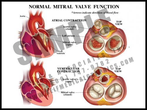
- Also known as the left atrioventricular valve, the mitral valve has 2 leaflets which are anchored to the ventricle floor by papillary muscles and chordae tendinae, as are the leaflets of the right atrioventricular valve (tricuspid valve).
- The aortic and pulmonary (pulmonic) valves are semilunar valves, and have thin cusps with thickened edges which seal during diastole (when the ventricles are relaxed and blood flows into the atria).
- The mitral valve prevents backflow from the left ventricle into the atrium; minor mitral valve prolapse or leak is usually clinically insignificant.
-
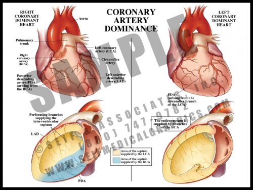
- Right heart dominance: the posterior portion of the interventricular septum is supplied by the posterior descending branch of the right coronary artery.
- Left heart dominance: the entire septum is supplied by branches of the left anterior descending artery; an obstruction in that vessel may lead to loss of the entire septum, an often fatal event. The posterior descending artery is derived from a branch of the circumflex artery instead of from the RCA.
-
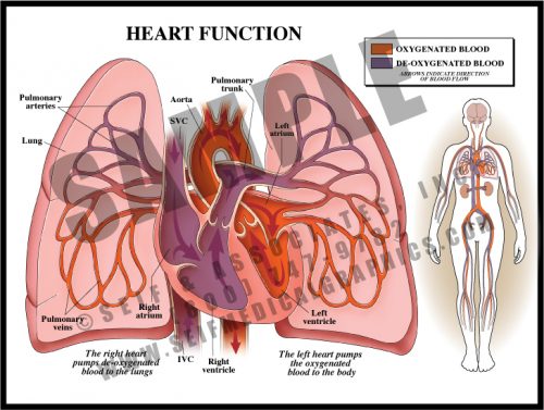
- The normal heart is really two separate pumps working in tandem; there is no connection between the right and left sides in the normal post-fetal heart.
- The right heart receives de-oxygenated blood from the body, moving it from the right atrium to the right ventricle to the lungs via the pulmonary artery. Carbon dioxide is released and oxygen is picked up in the lungs.
- The left heart receives oxygenated blood from the lungs, moving it from the left atrium to the left ventricle, and from there to the aorta, which distributes it to the rest of the body.
- The ventricles are thick muscular chambers which move blood with each contraction; the average left ventricle contracts with a force of 120 mmHg.