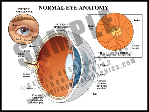
- The external eye has upper and lower lids which close over the globe to protect it. The sclera is the white of the eye, the colored portion is the iris, and the black opening in the middle of the iris is a hole known as the pupil. This is the only window in the body through which the nervous system can be seen directly.
- The anterior transparent media consists of the cornea, anterior chamber and lens; the posterior elements of the globe are covered with specialized nerve tissue, the retina.
- The optic nerve enters the eye posteriorly along with its own blood supply; this area is known as the optic disc. The macular area is where visual acuity is greatest.
-
-
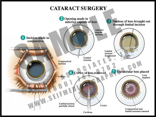
- The lens opacifies with age or after trauma. When vision is sufficiently affected, the cataract can be surgically removed and replaced with an artificial intraocular lens.
- The cornea is lifted from an incision in the blue-grey line surrounding the iris, and the anterior surface of the lens is opened. The nucleus of the lens is removed, leaving the posterior capsule of the lens in position.
- The intraocular lens is then placed within the capsule and fixed into position. Laser treatments are often needed post-operatively to clear the posterior capsule.
- This procedure is one of the safest and most common surgical procedures performed today, with a very low rate of complications.
-
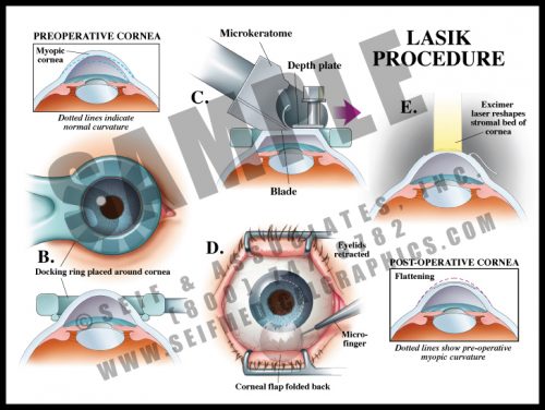
- LASIK (laser-assisted in situ keratomileusis) is a popular type of refractive surgery, or surgery performed to improve visual acuity.
- In LASIK, an incision is made to lift up a partial thickness of the cornea, using a very sharp, thin microtome.
- Once the flap is formed, the stroma of the cornea is sculpted with the laser, under computer control. Many of the newer LASIK systems can also accommodate for any eye movement during surgery, using a tracking program.
- This procedure has a very high success rate with relatively few complications.
-
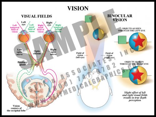
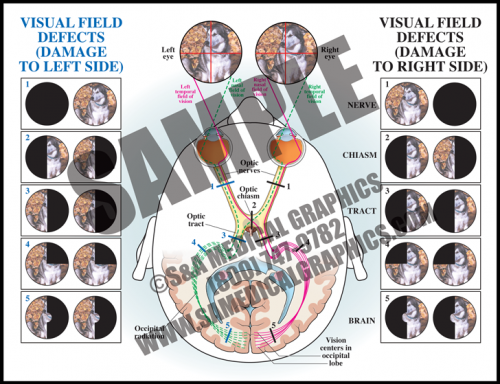
- The visual system is composed of specialized nerve fibers originating in the retina. They join to form the optic nerve (cranial nerve II) just behind the globe. The medial portions of the optic nerve cross over each other at the optic chiasm in front of the pituitary gland. As the nerve fibers travel posteriorly within the brain, they form the optic tracts terminating in the occipital lobe of the brain. By identifying visual field defect patterns, it is possible to determine an anatomical location of the source of visual loss.
- The visual fields of normally-aligned eyes overlap. Each eye sees objects from a slightly different angle, and the brain fuses these views. This binocular vision allows us to perceive depth and spatial relationships.
-
- A variety of retinal or more central pathologies can cause visual field deficits that are limited to particular regions of visual space.
- By identifying visual field defect patterns, it is possible to determine the anatomical location of the source of visual loss.
- Damage to the retina or one of the optic nerves before it reaches the optic chiasm results in a loss of vision that is limited to the eye of origin, while damage in the region of the optic chiasm or farther back in the brain will involve the visual fields of both eyes.
