This case involved a previously healthy 67-year-old male patient who presented with nearly fatal urosepsis from kidney stones. During this acute illness, he developed multisystem organ failure that required aggressive treatment. He survived the event, but sustained serious microvascular injury to his fingers and toes although his large vessels and pulses remained intact.
Unfortunately, all of the patient’s toes and several of his fingers developed gangrene. The medical team was able to salvage most of the fingers, but the toes required amputation after final demarcation. The lateral heels were gangrenous as well and the patient developed osteomyelitis. He was treated aggressively over the following months with home health care, multiple antibiotics, VAC treatments, and multiple debridements. The wounds waxed and waned, but ultimately the patient required bilateral below-the-knee amputations.
The plaintiff had several allegations, the first of which was that the damage to the patient’s heel was due to decubiti and not microvascular injury. They further alleged that the patient should have been referred to a specialist for a possible microvascular reconstruction and free muscle flap placement. The defense contended that this patient did not have heel decubiti; his initial illness had caused microvascular damage so that there were no good vessels remaining in his left heel. Further, the success rate of free flaps following heel osteomyelitis is very low. The medical team did everything they could to salvage this patient’s foot, but the microvascular injury was too severe and ultimately resulted in the amputations.
The plaintiff had several allegations, the first of which was that the damage to the patient’s heel was due to decubiti and not microvascular injury. They further alleged that the patient should have been referred to a specialist for a possible microvascular reconstruction and free muscle flap placement.
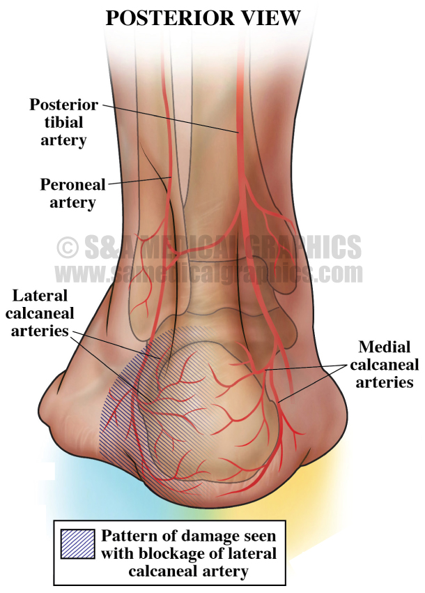
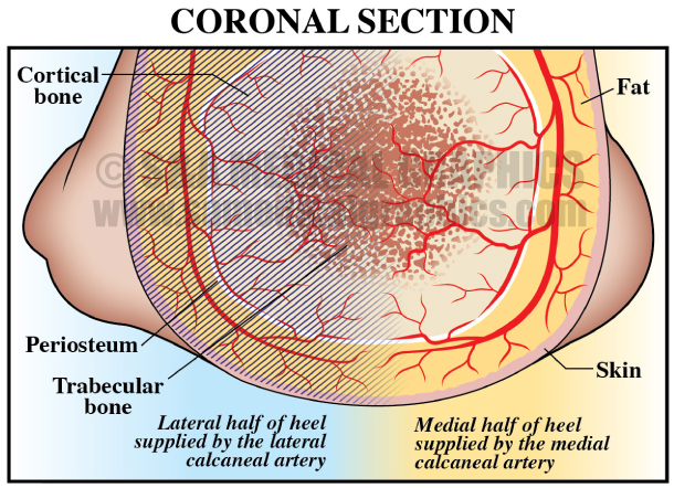
Since most of the issues in this case were closely related to the microvascular arterial supply of this patient’s feet, the defense first had to educate the jury on the anatomy of the blood supply to the foot and specifically, to the heel. The first exhibit of the defense’s visual strategy utilized areas of highlight and shading to simply explain to the jury which arteries supply the lateral versus medial heel. This simply demonstrated to the jury the pattern of damage that would be expected with damage to the lateral calcaneal artery compared to what would be expected with a heel decubiti. A corresponding exhibit showed the damage to this patient’s feet, thus allowing the jury to easily see that the patient’s pattern of damage was consistent with lateral calcaneal artery injury and did not match what would be expected for a heel decubiti.
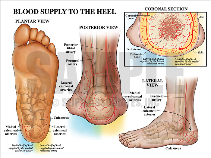
Exhibit 1 depicts the blood supply to the heel.
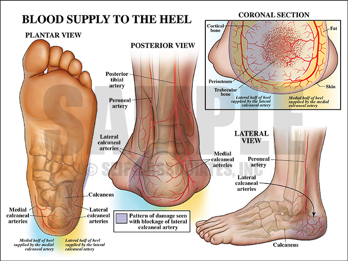
Exhibit 2 depicts pattern of damage with blockage of lateral calcaneal artery.
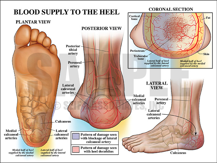
Exhibit 3 depicts the pattern of damage seen with heel decubitus.
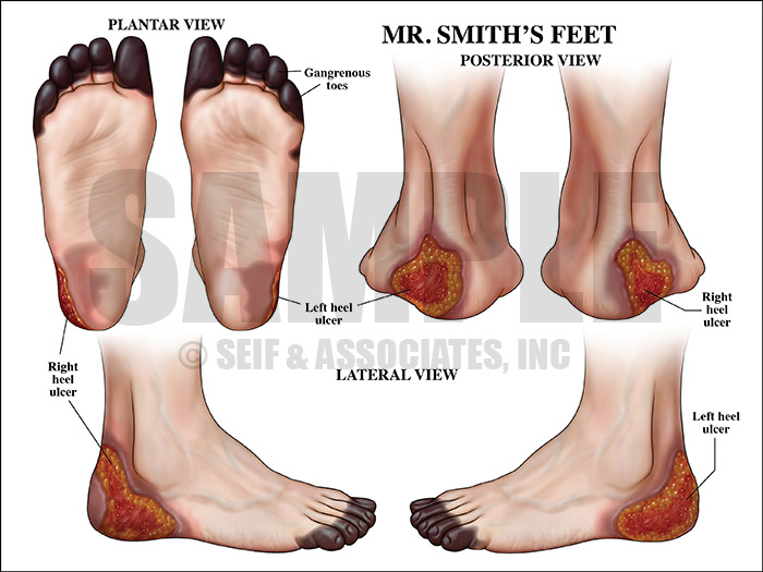
Exhibit 4 depicts extent of damage to patient’s feet.
With the next exhibit, the defense gave the jury a deeper understanding of the condition of osteomyelitis, showing how it occurs and why it is so difficult to manage.
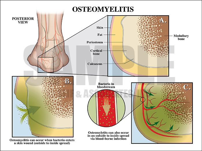
Exhibit 5 explains the condition of osteomyelitis.
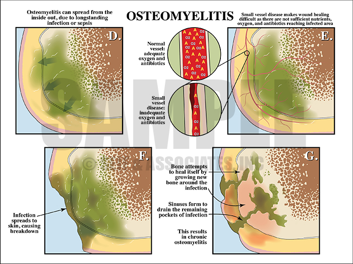
Exhibit 6 continues to explain the effects of osteomyelitis.
In order to counter the plaintiff’s allegation that the patient should have been referred to a specialist for a free flap closure of the defect, we created an exhibit to explain free flap microvascular anastomses to the jury. The exhibit also highlighted the high rate of amputation in patients with heel ulcers and osteomyelitis who undergo this procedure. This exhibit was designed to draw the jury’s attention to the fact that even if this patient had undergone this invasive procedure, there is no guarantee that he would have avoided amputation.

Exhibit 7 depicts a free flap microvascular anastomosis procedure.
Lastly, the defense sought to visually explain to the jury why VAC therapy was a good option for this patient. By simply showing how this system heals wounds, it was the defense’s hope that the jury would see why this treatment option was valid and certainly did not constitute negligence.
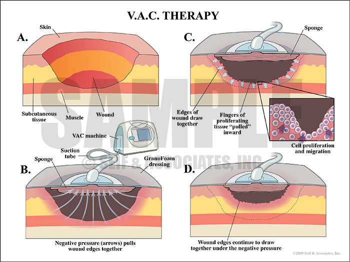
Exhibit 8 depicts how VAC therapy heals wounds.
—Editorial contributed by Emily Ullo Steigler, MS, CMI
—Illustrations contributed by Robert Edwards, MS, CMI