Generally speaking, gynecologic surgery accounts for over 50% of all ureteral injuries resulting from operations. Hysterectomy of any type is usually a safe procedure, but any surgery has risks, such as blood loss, blood clots, infection, damage to surrounding organs, and reactions to anesthesia. Surgery involving the organs in the pelvis puts the surrounding organs at risk of injury as well.
Bladder, ureter, and bowel injuries are relatively common and known complications of hysterectomy, whether total abdominal, vaginal, laparoscopic, or laparoscopically-assisted. Injuries can be due to stapling/sutures, thermal injury, or aberrant anatomy. Subsequently, plaintiff claims of negligence as a result of these types of injuries are not uncommon in the realm of medical malpractice negligence. The following case study examines the visual strategy of the defense in an interesting case of ureteral injury after vaginal hysterectomy.
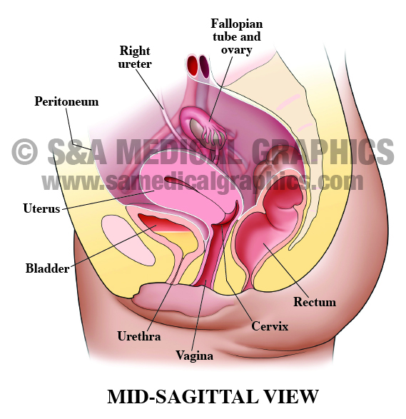
Mid-sagittal view of female pelvis.
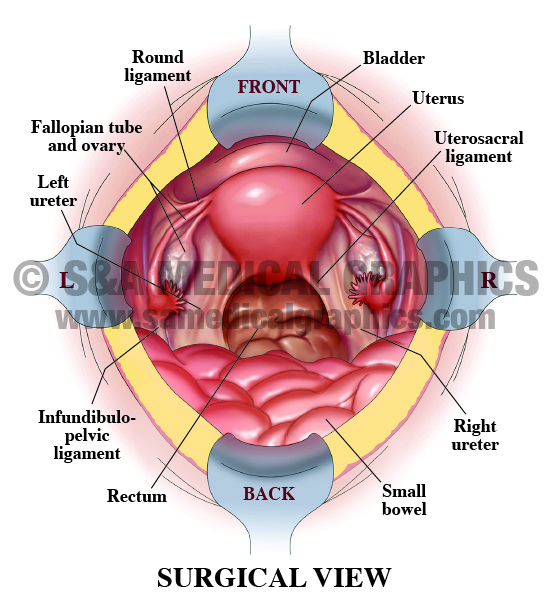
Surgical view of female pelvis during
hysterectomy procedure.
The female plaintiff presented to the defendant for a vaginal hysterectomy. There were no unusual findings and the procedure was without complication. At the conclusion of the procedure, a Foley was placed and the plaintiff was voiding well with clear urine. Postoperatively, the plaintiff complained of right-sided hip pain, but it was generally felt to be discomfort due to positioning. The plaintiff was discharged on the first postoperative day voiding well with clear urine. Two days later, the plaintiff returned to the defendant complaining of pain involving her right flank. She had a history of kidney stones, so she was sent for consultation. Two days postoperatively, a cystoscopy showed a right-sided ureteral obstruction with hydronephrosis. Ultimately, the plaintiff underwent a re-implantation of the ureter and insertion of the right-sided nephrostomy tube.
The plaintiff alleged that the defendant tied the ureter off with a suture during the vaginal hysterectomy procedure. The defense contended that the ureteral obstruction was caused by kinking of the ureter and/or by post-operative swelling of the surrounding tissues. While unfortunate, they argued that ureteral injury is a known complication of this procedure.
In order to visually present the defense strategy in court, the plan was to first orient the jury to the anatomy of the female pelvis involved in this case (Exhibit 1). This gave the jury a frame of reference for the female anatomy as well as highlighting the spatial relationship between the uterus/ vagina, the bowel, and the ureter.
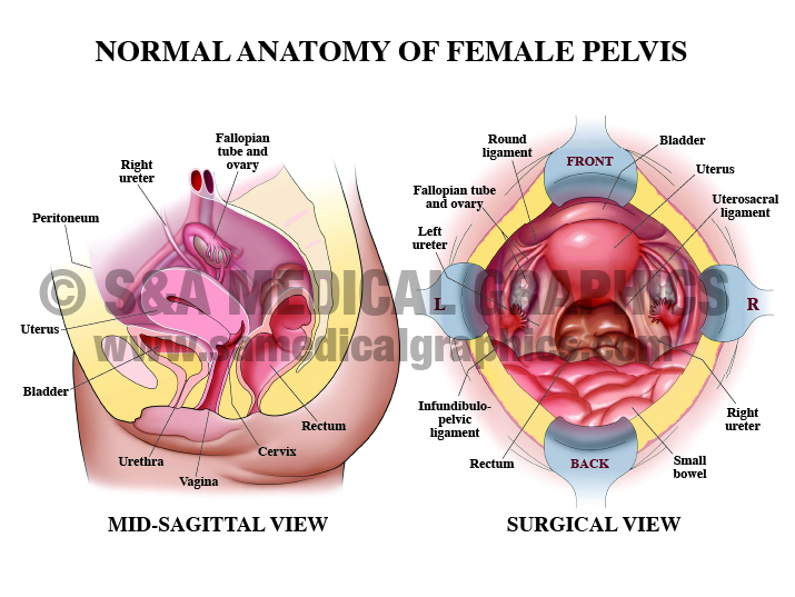
Exhibit 1 depicts the normal anatomy of a female pelvis.
The next step in the visual strategy was to provide a visual reference of the plaintiff’s surgical procedure in order to help the defendant explain what he did during the procedure (Exhibit 2).
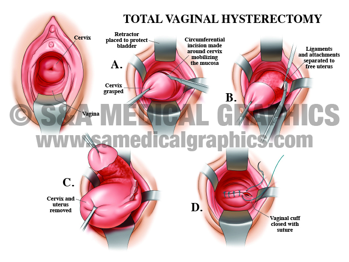
Exhibit 2 depicts the surgical steps of a total vaginal hysterectomy.
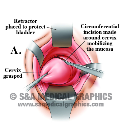
Close-up of step A showing incision.
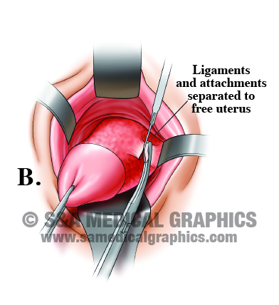
Close-up of step B showing ligaments
removed around uterus.
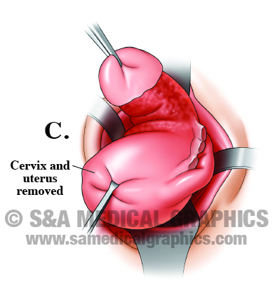
Close-up of step C showing the cervix and
uterus being removed.
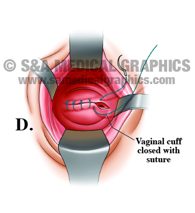
Close-up of step D showing the vaginal cuff
being closed with sutures.
The final portion of the visual strategy was to show the jury the likely mechanism of injury in this case (Exhibit 3). By showing how the closure of the vaginal cuff could put tension on the right side of the cuff, causing the right ureter to kink, blocking the flow of urine. It would also demonstrate how the swelling and inflammation of the surrounding tissues could contribute to the kinking and blockage.
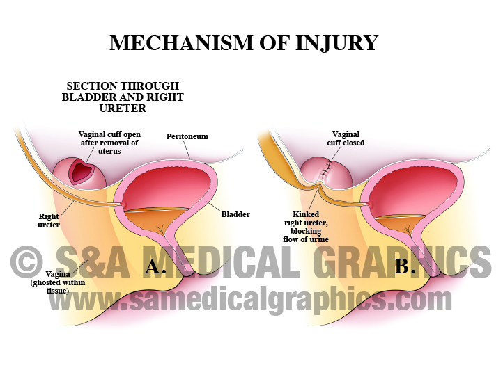
Exhibit 3 depicts the mechanism of injury from hysterectomy procedure.
The defense found these exhibits to be helpful in creating a baseline of medical knowledge for the jury and allowing their client and experts to explain their theories of alternative causation. Ultimately, the defense received a favorable outcome for their client in this case.
—Editorial contributed by Emily Ullo Steigler, MS, CMI
—Illustrations contributed by Jennifer C. Webb, MS
